
ECHINOCOCCOSIS: COMPLICATED HYDATID CYST OF THE LIVER
Keywords:
hydatid cyst, echinococcosis, complication, diagnostic search, computed tomography (CT), magnetic resonance imaging (MRI), ultrasound investigation (USI)
Abstract
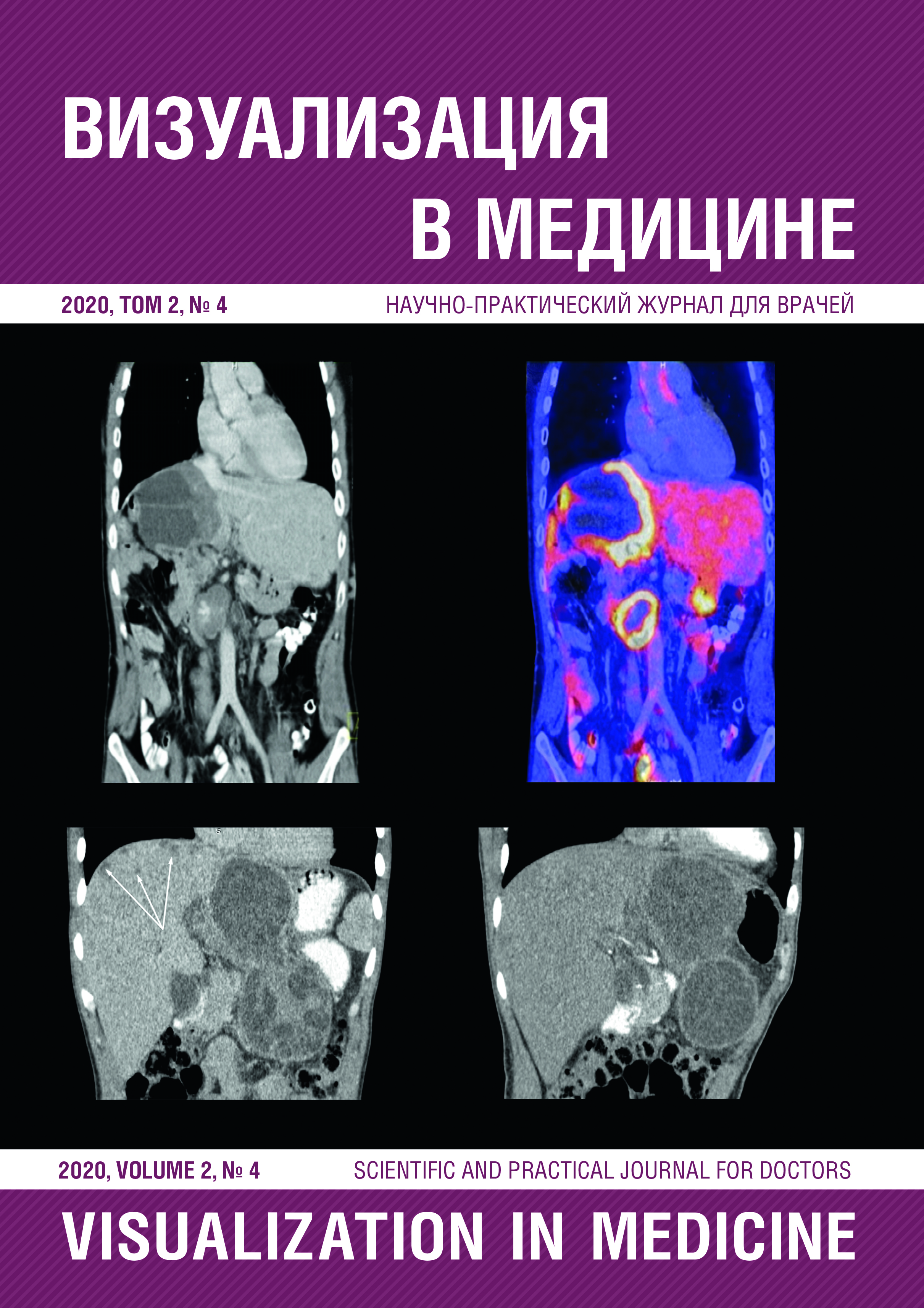
Nowadays, despite group and individual prevention measures which may be taken, echinococcosis still remains a dangerous zoonosis whose diagnosis is difficult to establish due to the non specific clinical picture with a long term period of asymptomatic course. This clinical case brightly illustrates the capabilities to visualize the manifestations of different stages of one patient’s disease using the computed tomography (CT), as well as the significance of the method for determining the tactics of further treatment. The article provides examples of specific radiological symptoms of hydatid cyst and its complications which allow the attending physician to choose the right way of diagnostic search.
Published
2021-04-14
How to Cite
Пивсаева, В. М., Пивсаев, А. В., & Батынков, С. П. (2021). ECHINOCOCCOSIS: COMPLICATED HYDATID CYST OF THE LIVER. Visualization in Medicine, 2(4), 28-38. Retrieved from https://ojs3.gpmu.org/index.php/visual-med/article/view/2597
Issue
Section
Статьи

