THE ROLE OF ARTIFICIAL INTELLIGENCE IN THE EVALUATION OF X-RAY COMPUTED TOMOGRAPHY DATA IN PATIENTS WITH INTERSTITIAL LUNG DISEASES IN THE COVID-19 PANDEMIC
Abstract
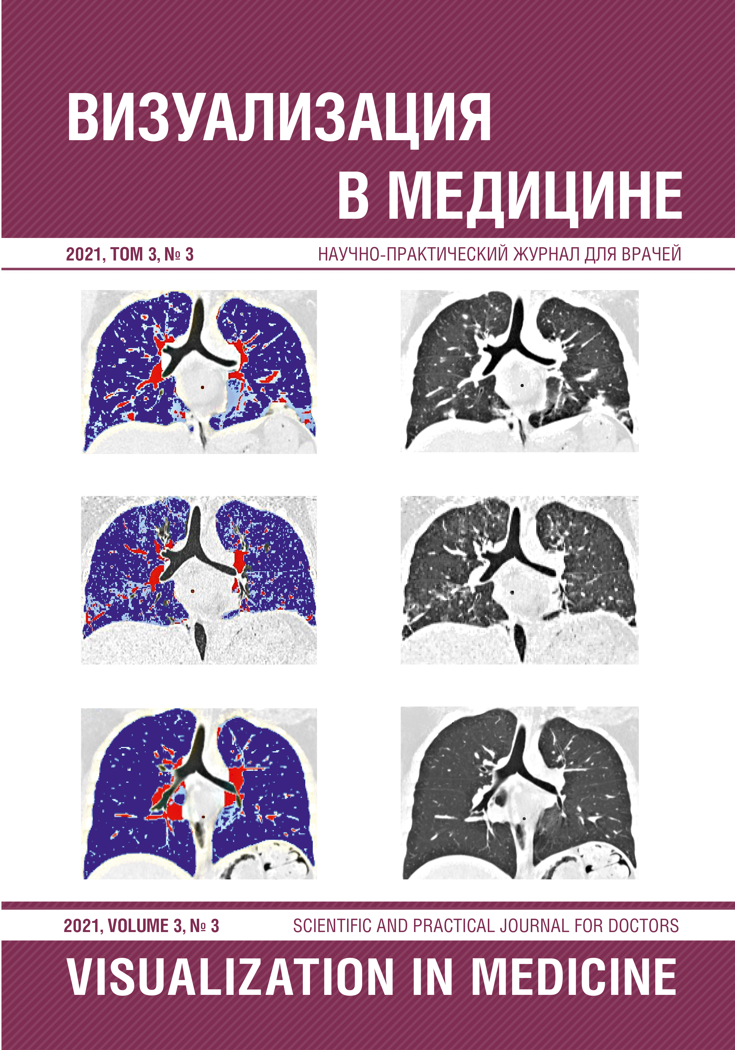
The purpose of the study. To evaluate the possibility of artificial intelligence programs in the evaluation of X-ray computed tomography data in patients with postcovid changes, other interstitial lung diseases (ISL), as well as their combination. Materials and methods. The data of 16 patients with ISL, who had a new coronavirus infection, observed from March 2020 to March 2021 at the Pavlov PSPbSMU were analyzed. Patients with ISL were divided into 2 groups: patients with progressive pulmonary fibrosis (ILF - 8, LF syndrome with DBST - 2, toxicoallergic alveolitis - 1) and cystic lung lesion (LAM - 3, GC - 2). In all patients, the diagnosis of ISL was confirmed morphologically, COVID-19 - by PCR. At the time of the addition of a new coronavirus infection, the acute nature of the course of the disease was noted with the presence of shortness of breath (up to shortness of breath at rest), the addition or increase in dry cough, an increase in body temperature, an increase in acute blood parameters and radiation signs of acute interstitial pneumonia on CT. The average age of patients was 54.3 ± 10.1 years (w/m - 7/9). All patients underwent VRKT, if possible, a comprehensive functional study of external respiration (CPIVD) and echocardiography. In all patients, a postprocessor image analysis was performed with an assessment of the volume of lung tissue lesion using the «density mask» programs. Data were compared with the assessment of the lesion volume in patients with only COVID-19 pulmonary pathology, a combination of COVID-19 with ISL and independent lung damage in progressive pulmonary fibrosis and cystic bullous processes. Results. The analysis of the results of the radiation study revealed the undoubted advantages of artificial intelligence in helping to assess the volume of lung damage, allowing: 1) to estimate the exact percentage of lung tissue damage using programs for isolating densities characteristic of COVID-19 lung pathology; 2) in patients with cystic bullous transformation of the lungs, the «density masks» programs allowed to simultaneously assess both the % of lung tissue damage associated with COVID-19 and the degree of prevalence of the main process; summation of these indicators made it possible to estimate the preserved reserve of lung volumes; 3) in patients with progressive pulmonary fibrosis, COVID-19 caused an increase in the extent and deterioration of the qualitative indicators of the fibrous process. In terms of density characteristics, fibrous changes did not differ from COVID-19 indicators and could be used to assess the progression of pulmonary fibrosis; 4) the obtained data allow the use of a program calculation with the use of density characteristics of interstitial lung disease to assess the fibrosis progression of another origin (idiopathic, with diffuse connective tissue diseases, after suffering a toxic allergic and exogenous allergic alveolitis); 5) of a similar character calculation of volumes using the program «density mask» can be used for evaluation of the severity of cystic changes in lymphangioleiomyomatosis (LAM) and histiocytosis X (GC). Conclusions. The accumulation of experience in clinical and radiation examination of patients with a combination of interstitial lung diseases and COVID-19 has allowed us to develop a radiation algorithm for their qualitative and quantitative assessment, which is important for determining therapeutic tactics. The developed model for calculating the affected lung volume can be applied to progressive pulmonary fibrosis and cystic bullous processes in the lungs.