КЛИНИКО-РЕНТГЕНОЛОГИЧЕСКИЕ ОСОБЕННОСТИ ПЕПТИЧЕСКИХ ЯЗВ АНАСТОМОЗА ПРИ СИНДРОМЕ ЗОЛЛИНГЕРА-ЭЛЛИСОНА
Аннотация
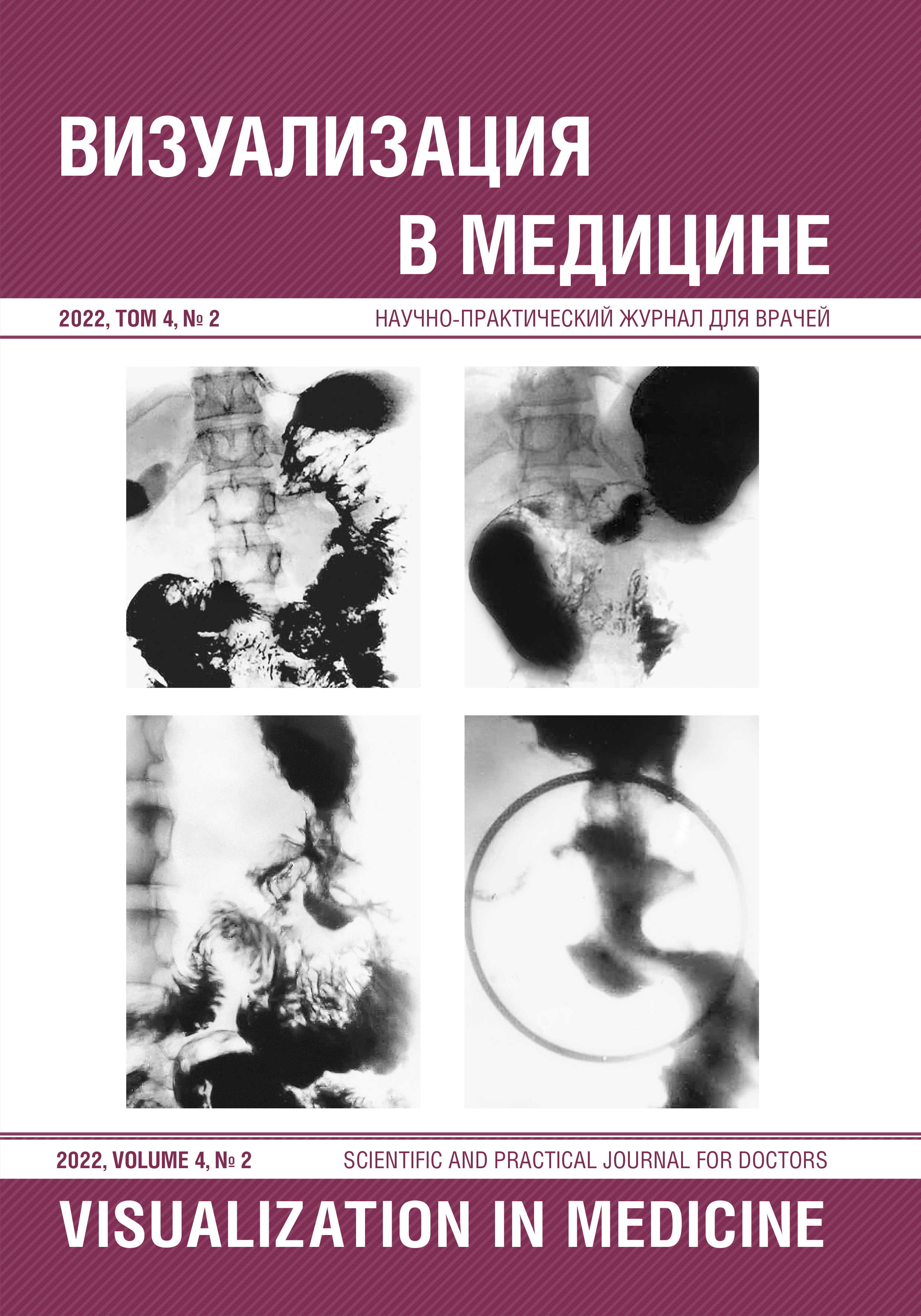
Ведущее значение в распознавании пептических язв анастомоза имеет рентгенологический метод исследования. Несмотря на то что рентгенологические признаки пептических язв тощей кишки достаточно подробно описаны, определенные трудности представляет трактовка полученных данных из -за грубой деформации исследуемого отдела, а также различных тенеобразований, симулирующих симптомы язвенной ниши. Именно поэтому целью работы было выявление клинико -рентгенологических паттернов при синдроме Золлингера-Эллисона. Результаты. Проанализированы результаты обследования 24 пациентов с синдромом Золлингера-Эллисона. У всех пациентов присутствовал болевой симптом. В 58% случаев это были тупые боли, в 42% случаев - острые. У 84% пациентов локализация боли соответствовала эпигастральной области. Рвота выявлена только в 29% случаев. Светлый промежуток отсутствовал у 9 пациентов, у 15 пациентов он длился от 7 месяцев до 5 лет. Свободная соляная кислота в пределах 40-60 титр. единиц наблюдалась у 8 пациентов, в остальных случаях в пределах 61-150 титр. единиц. Желудочно -кишечные кровотечения встречались у 14 пациентов. 22 пациента перенесли оперативные вмешательства по поводу рецидива пептической язвы тощей кишки, причем 10 - резекцию, и 12 - стволовую ваготомию. Эндоскопия выполнена 11 пациентам, язва выявлена у 6. При рентгенологическом исследовании язвенная ниша была выявлена у 22 (92%) пациентов, причем в одном случае были обнаружены три язвенные ниши. У большинства пациентов (20 - 83%) были выявлены ниши более 2 см в диаметре и только у двух - ниши были размерами 1,0 и 1,5 см. Рубцово -язвенная деформация встречалась у всех больных, причем наиболее часто (19 случаев) - I и II степени. Заключение. Таким образом, у пациентов с синдромом Золлингера-Эллисона наряду с типичными клиническими его проявлениями встречались случаи нетипичного течения - слабо выраженный болевой симптом, отсутствие кровотечений, низкие цифры кислотности. Размеры язв большей частью были большими, однако встречались случаи, когда имелись небольшие язвы (1,0-1,5 см в диаметре). Гигантские язвы были выявлены у пациентов с низкими цифрами кислотности. Подобные атипичные случаи могут затруднять распознавание синдрома Золлингера-Эллисона.