KIDNEY DAMAGE IN LEUKEMIA (CLINICAL CASE)
Abstract
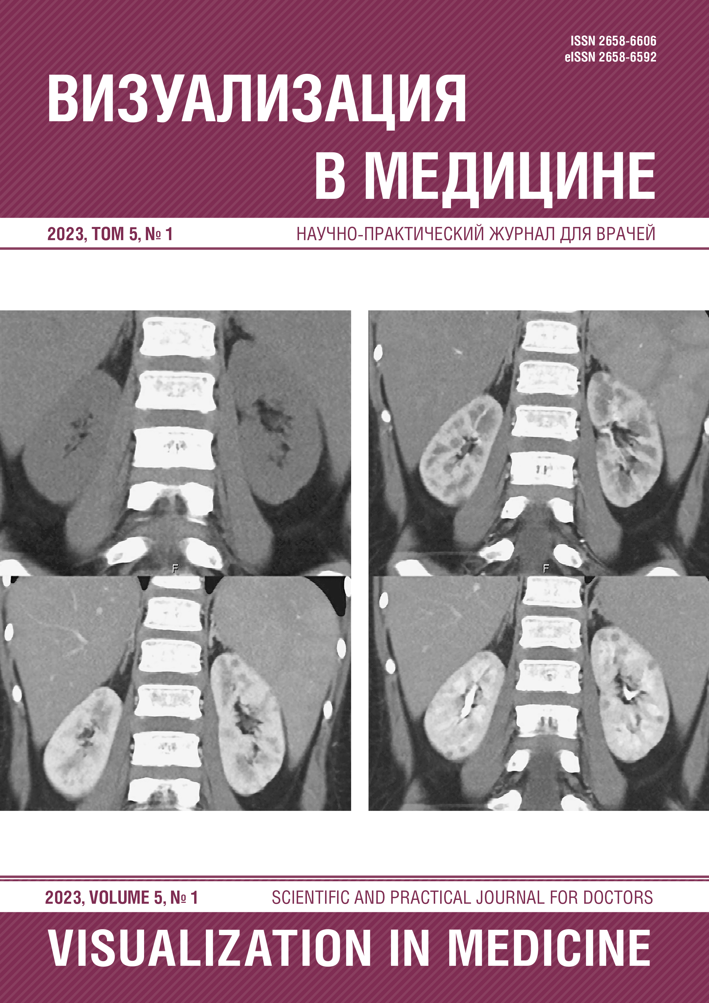
Leukemia is a group of diseases of the blood system that affects the hematopoietic cells of the bone marrow at different stages of their maturation by mutation of cellular DNA, as a result of which the cell acquires a number of new characteristics, including loss of the ability to further differentiate and a tendency to uncontrolled proliferation, which causes the classic clinical picture of this pathology. Acute lymphoblastic leukemia (ALL) ranks first in the structure of oncological morbidity among children. Most cases of ALL in pediatric practice are diagnosed at the age of 2 to 5 years (75% of patients). Clinical manifestations of ALL in children are diverse and are caused by leukemic infiltration and impaired function of the affected organs. Infiltration of bone marrow tumor cells leads to the development of anemia, thrombocytopenia, leukocytosis or leucopenia. In the case of a prolonged course of ALL, in the absence of treatment, an extramedullary lesion occurs due to infiltration by leukemic cells of internal organs (liver, spleen, kidneys, testicles, central nervous system), lymph nodes, bones, skin. The gold standard for the diagnosis of ALL is morphological, molecular genetic, cytogenetic examination, as well as flow cytometry of bone marrow cells. In order to identify the foci of extramedullary lesions in ALL, imaging research methods are used. Kidney damage in ALL manifests as nephromegaly due to infiltration by leukemic cells. According to the literature, microscopic infiltration of the kidneys occurs in 7–42% of cases, ALL is more often recorded in advanced stages of the disease. This article presents a clinical case of bilateral kidney damage in ALL with a CT picture that required a differential diagnosis with polycystic kidney disease. The purpose of this article is to demonstrate the features and capabilities of radiation imaging of the pathology of the blood system, kidneys and bone and joint apparatus in leukemia.