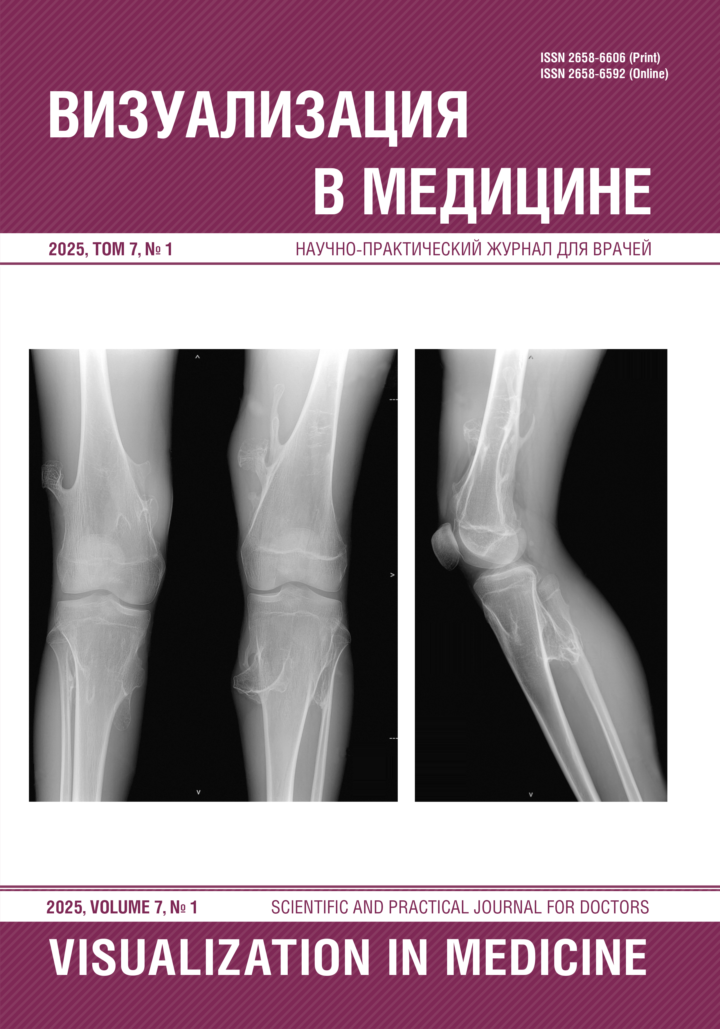
MODERN VIEW ON HEREDITARY MULTIPLE EXOSTOSES (LITERATURE REVIEW, CASE REPORT)
Abstract
Hereditary multiple exostoses (HME) is a relatively rare hereditary disorder characterized by the formation of multiple osteochondromas. HME has an autosomal dominant inheritance pattern and is characterized by variable expressivity, which leads to significant differences in clinical manifestations. Most often, HME is caused by mutations in the exostosin-1 (EXT-1) and exostosin-2 (EXT-2) genes, which play an important role in the synthesis of heparan sulfate (HS), which is involved in the regulation of bone and cartilage tissue formation. A defect in the synthesis of HS leads to disruption of normal bone growth, which is manifested by the development of osteochondromas — benign osteochondral formations that occur mainly in the area of long tubular bones, especially in the metaphyseal zones. Although HME may be asymptomatic, patients with this disease experience a wide range of clinical manifestations. Symptoms range from localized pain caused by the pressure of the tumor on surrounding tissues to severe bone deformities and functional disorders. Skeletal deformities may lead to shortening of the limbs, poor posture, limited joint mobility, and compression of vascular and nerve structures. One of the most dangerous complications of HME is the malignant transformation of osteochondroma into secondary chondrosarcoma (1–5% cases). The therapeutic approach to HME consists of dynamic observation and surgical resection if indicated. In recent years, studies have been conducted on the possible use of targeted therapy aimed at correcting impaired heparan sulfate metabolism, but the clinical application of such methods is still at the study stage. The aim of this article is to review updated data regarding the epidemiology, pathogenesis, clinical presentation, imaging modalities, treatment options, and prognosis of HME.
References
Гайдук И.М., Баирова С.В., Полищук Т.В., Булычева В.И., Ревнова М.О., Сахно Л.В., Колтунцева И.В., Мишкина Т.В., Орел В.И., Ким А.В., Рослова З.А. Организация медико-социальной помощи подросткам в современных условиях. Медицина и организация здравоохранения. 2021;6(3):84–95.
Косинская Н.С. Наследственные заболевания скелета: классификация, патогенез и клинические проявления. М.: Медицина; 1968.
Орел В.И., Середа В.М., Ким А.В., Шарафутдинова Л.Л., Беженар С.И., Булдакова Т.И., Рослова З.А., Орел В.В., Гурьева Н.А. Здоровье детей Санкт-Петербурга. Педиатр. 2017;8(1):112–119. DOI: 10.17816/PED81112-119.
Потапчук А.А., Дидур М.Д. Осанка и физическое развитие детей. Программы диагностики и коррекции нарушений. СПб.; 2001.
Ahmed A.R., Tan T.S., Unni K.K., Collins M.S., Wenger D.E., Sim F.H. Secondary chondrosarcoma in osteochondroma: report of 107 patients. Clin Orthop Relat Res. 2003;411:193–206. DOI: 10.1097/01.blo.0000069888.31220.2b.
Alyas F., James S.L., Davies A.M., Saifuddin A. The role of MR imaging in the diagnostic characterisation of appendicular bone tumours and tumour-like conditions. Eur Radiol. 2007;17(10):2675–86. DOI: 10.1007/s00330-007-0597-y.
Aoki J., Watanabe H., Shinozaki T., Tokunaga M., Inoue T., Endo K. FDG-PET in differential diagnosis and grading of chondrosarcomas. J Comput Assist Tomogr. 1999;23(4):603–8. DOI: 10.1097/00004728-199907000-00022.
Bovée J.V. Multiple osteochondromas. Orphanet J Rare Dis. 2008;3:3. DOI: 10.1186/1750-1172-3-3.
Brien E.W., Mirra J.M., Luck JV. Jr. Benign and malignant cartilage tumors of bone and joint: their anatomic and theoretical basis with an emphasis on radiology, pathology and clinical biology. II. Juxtacortical cartilage tumors. Skeletal Radiol. 1999;28(1):1–20. DOI: 10.1007/s002560050466.
D’Arienzo A., Andreani L., Sacchetti F., Colangeli S., Capanna R. Hereditary Multiple Exostoses: Current Insights. Orthop Res Rev. 2019;11:199–211. DOI: 10.2147/ORR.S183979.
Douis H., Saifuddin A. The imaging of cartilaginous bone tumours. I. Benign lesions. Skeletal Radiol. 2012;41(10):1195–212. DOI: 10.1007/s00256-012-1427-0.
Francannet C., Cohen-Tanugi A., Le Merrer M., Munnich A., Bonaventure J., Legeai-Mallet L. Genotype-phenotype correlation in hereditary multiple exostoses. J Med Genet. 2001;38(7):430–4. DOI: 10.1136/jmg.38.7.430.
Garcia R.A., Inwards C.Y., Unni K.K. Benign bone tumors — recent developments. Semin Diagn Pathol. 2011;28(1):73–85. DOI: 10.1053/j.semdp.2011.02.013.
Gavanier M., Blum A. Imaging of benign complications of exostoses of the shoulder, pelvic girdles and appendicular skeleton. Diagn Interv Imaging. 2017;98(1):21–28. DOI: 10.1016/j.diii.2015.11.021.
Göçmen S., Topuz A.K., Atabey C., Şimşek H., Keklikçi K., Rodop O. Peripheral nerve injuries due to osteochondromas: analysis of 20 cases and review of the literature. J Neurosurg. 2014;120(5):1105–12. DOI: 10.3171/2013.11.JNS13310.
Hakim D.N., Pelly T., Kulendran M., Caris J.A. Benign tumours of the bone: A review. J Bone Oncol. 2015;4(2):37–41. DOI: 10.1016/j.jbo.2015.02.001.
Hameetman L., Bovée J.V., Taminiau A.H., Kroon H.M., Hogendoorn P.C. Multiple osteochondromas: clinicopathological and genetic spectrum and suggestions for clinical management. Hered Cancer Clin Pract. 2004;2(4):161–173. DOI: 10.1186/1897-4287-2-4-161.
Herget G.W., Kontny U., Saueressig U., Baumhoer D., Hauschild O., Elger T., Südkamp N.P., Uhl M. Osteochondrom und multiple Osteochondrome: Empfehlungen zur Diagnostik und Vorsorge unter besonderer Berücksichtigung des Auftretens sekundärer Chondrosarkome. Radiologe. 2013;53(12):1125–36. (In German). DOI: 10.1007/s00117-013-2571-9.
Khosla A., Parry R.L. Costal osteochondroma causing pneumothorax in an adolescent: a case report and review of the literature. J Pediatr Surg. 2010;45(11):2250–3. DOI: 10.1016/j.jpedsurg.2010.06.045.
Kitsoulis P., Galani V., Stefanaki K., Paraskevas G., Karatzias G., Agnantis N.J., Bai M. Osteochondromas: review of the clinical, radiological and pathological features. In Vivo. 2008;22(5):633–46.
Legeai-Mallet L., Munnich A., Maroteaux P., Le Merrer M. Incomplete penetrance and expressivity skewing in hereditary multiple exostoses. Clin Genet. 1997;52(1):12–6. DOI: 10.1111/j.1399-0004.1997.tb02508.x.
Motamedi K., Seeger L.L. Benign bone tumors. Radiol Clin North Am. 2011;49(6):1115–34. DOI: 10.1016/j.rcl.2011.07.002.
Murphey M.D., Choi J.J., Kransdorf M.J., Flemming D.J., Gannon F.H. Imaging of osteochondroma: variants and complications with radiologic-pathologic correlation. Radiographics. 2000;
(5):1407–34. DOI: 10.1148/radiographics.20.5.g00se171407.
Pacifici M. Hereditary Multiple Exostoses: New Insights into Pathogenesis, Clinical Complications, and Potential Treatments. Curr Osteoporos Rep. 2017;15(3):142–152. DOI: 10.1007/s11914-017-0355-2.
Pontes ÍCM., Leão R.V., Lobo CFT., Paula V.T., Yamachira V.S., Baptista A.M., Helito PVP. Imaging of solitary and multiple osteochondromas: From head to toe — A review. Clin Imaging. 2023;103:109989. DOI: 10.1016/j.clinimag.2023.109989.
Ryckx A., Somers J.F., Allaert L. Hereditary multiple exostosis. Acta Orthop Belg. 2013;79(6):597–607.
Sakata T., Mogi K., Sakurai M., Nomura A., Fujii M., Takahara Y. Popliteal Artery Pseudoaneurysm Caused by Osteochondroma. Ann Vasc Surg. 2017;43:313.e5–313.e7. DOI: 10.1016/j.avsg.2017.04.003.
Sánchez-Rodríguez V., Medina-Romero F., Gómez Rodríguez-Bethencourt M.Á., González Díaz M.A., González Soto M.J., Alarcó Hernández R. Value of the bone scintigraphy in multiple osteochrondromatosis with sarcomatous degeneration. Rev Esp Med Nucl Imagen Mol. 2012;31(5):270–4. (In English, Spanish). DOI: 10.1016/j.remn.2011.10.011.
Tepelenis K., Papathanakos G., Kitsouli A., Troupis T., Barbouti A., Vlachos K., Kanavaros P., Kitsoulis P. Osteochondromas: An Updated Review of Epidemiology, Pathogenesis, Clinical Presentation, Radiological Features and Treatment Options. In Vivo. 2021;35(2):681–691. DOI: 10.21873/invivo.12308.
Williams J.A., Kondo N., Okabe T., Takeshita N., Pilchak D.M., Koyama E., Ochiai T., Jensen D., Chu M.L., Kane M.A., Napoli J.L., Enomoto-Iwamoto M., Ghyselinck N., Chambon P., Pacifici M., Iwamoto M. Retinoic acid receptors are required for skeletal growth, matrix homeostasis and growth plate function in postnatal mouse. Dev Biol. 2009;328(2):315–27. DOI: 10.1016/j.ydbio.2009.01.031.
Wootton-Gorges S.L. MR imaging of primary bone tumors and tumor-like conditions in children. Magn Reson Imaging Clin N Am. 2009;17(3):469–87. DOI: 10.1016/j.mric.2009.03.010.
Xing S.G., Mao T. Surgical excision of enchondromas and osteochondromas in the hand under local anaesthesia without tourniquet. J Hand Surg Eur Vol. 2019;44(7):745–747. DOI: 10.1177/1753193419845254.


